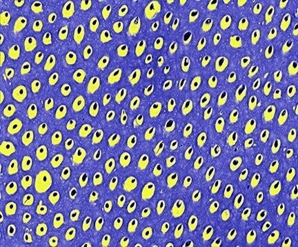
Zebra Dentine
The upper molar root dentine of a southern African zebra (Equus burchelli) is observed by backscattered electron imaging in the scanning electron microscope. The image derives from the polished cut surface of a tooth sectioned through its center. The image is called a “density-dependent” image; black represents hollow tubules (i.e. no dentine), blue is least densely mineralized (i.e. relatively less hard dentine), and yellow is most densely mineralized (i.e. relatively more hard dentine). Each tubule actually represents a tube associated with one long dentine cell process in life. The number of tubules and the proportion of yellow to blue may characterize certain species and relate to their feeding habits.
This research concerns interests in the skeletal and conservation biology of African mammals.Information obtained from our imaging research is expected to highlight the effects of environmental change, disease, and possibly the stresses related to living in protected game reserves (e.g. overcrowding and tourism). The results are also expected to be useful for environmental reconstruction of sites of importance to ancient human life.
Original width 265 μm

