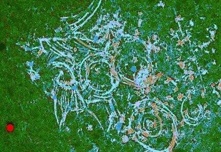
Lucy Fiber Orientation
Image of bone microanatomy by portable confocal scanning optical microscopy. A novel portable Nipkow disk-based confocal microscope invented and patented by the artist was employed in the imaging of femoral bone from the famous “Lucy” discovered from fossil bearing deposits at Hadar, Ethiopia, approximately 3.0 m.y old.
This image provides information about the degree of orientation of the collagen fibers within the bone, which in turn can tell us about the ability of the tissue to resist different kinds of mechanical stresses encountered in everyday life; green is collagen perpendicular with the plane of the screen, and light blue represents collagen parallel with the screen. Comparing the organization of bone tissue between Lucy, belonging to the species Australopithecus afarensis, with other species of early human, can help us to understand more about how bone structure and function has varied over human evolutionary time.
Original width 0.6 mm

