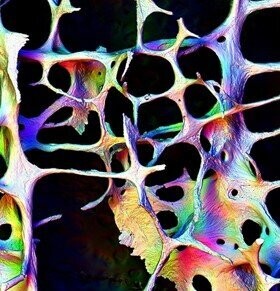
Human Trabecular Bone 1 & 2
Top: Spongy (trabecular) bone from the lumbar vertebra of an 89-year-old female is observed by backscattered electron imaging in the scanning electron microscope (SEM).Color hue shows the spatial orientation (direction in which it is facing) and color intensity shows the slope of the surface.Eleven levels (in-focus planes at consecutive depths) of 250 microns each were recorded separately to provide an image in good focus at all depths.The world’s first in-focus multi-level SEM image is presented here.This image, and the technique employed to produce it, allows a better discrimination of bone surface activity than has been achieved before.In this elderly female the beams of bone making up the inner architecture of the vertebra are significantly thinned compared to pre-menopausal woman.The novel imaging methods portrayed here affords a new perspective on osteoporosis.
Original width 4.45 mm
Bottom: Scanning electron micrograph of vertical slice of cancellous bone from a fourth lumbar (L4) vertebral body from an elderly female. Composite made from 36 backscattered electron images, 12 focus levels at 250 micron vertical separation.At each focus level, 4 images were recorded with separate backscattered electron detector sectors. Three of these images were combined by assigning the grey level image to one of three RGB colour channels. This gives the pleasing back-lit effect with subtle colour tones.All in focus images makes it possible to see all structural detail over all surfaces over a large depth range.
Original width 4 mm
Images courtesy of Professor Alan Boyde, Queen Mary University of London.


