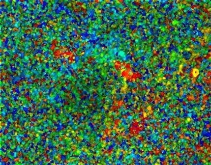
Atapuerca Cave Attack 1
Image of bone microanatomy by portable confocal scanning optical microscopy. A novel portable Nipkow disk-based confocal microscope invented and patented by the artist was employed in the imaging of bone from a bear skeleton discovered from human fossil bearing deposits at Atapuerca, Spain, approximately 0.4-0.7 m.y old.
Bones from Atapuerca have been severely affected by bacterial attack during fossilization, eliminating much of the internal microanatomy, but leaving the external macroanatomy in perfect condition. In order to visualize any remaining microanatomy it is necessary to use the autofluorescence potential of bone. Thus, instead of using normal white light, the microscope is configured to image the fluorescence of bone when using ultraviolet light. The image obtained here is a 3D image of bacterial attack, the colors depicting damage at various levels from top (red) to bottom (blue). The colors are very mixed up because of the degree and nature of the attack.
Original width 700 μm

