3D Bone Structure
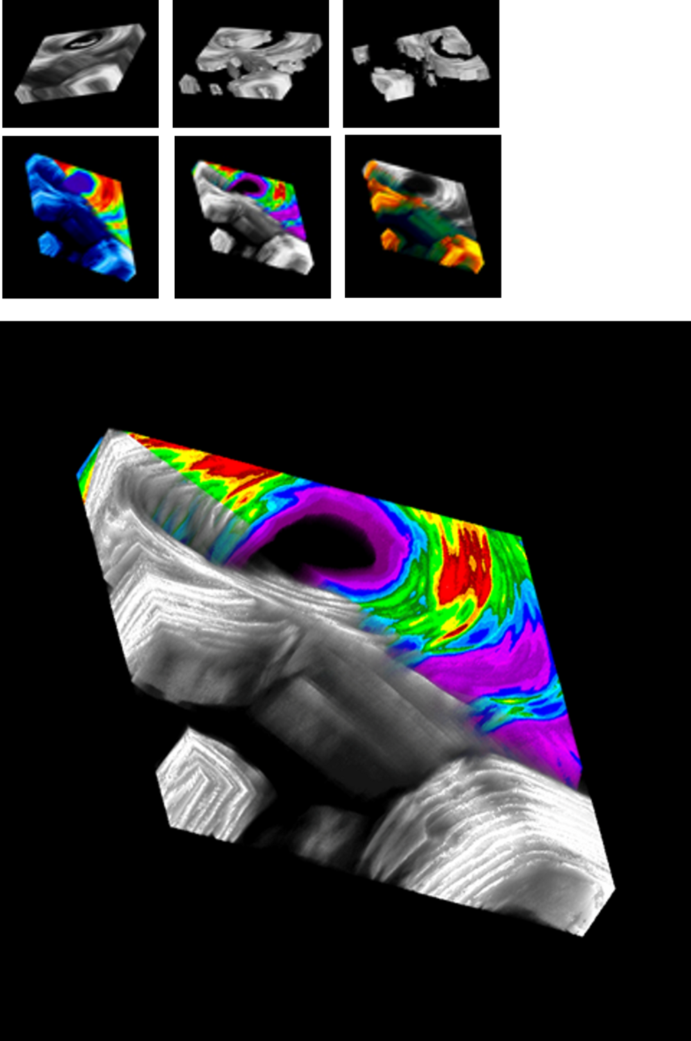
3D image of a blood vessel (top and center) contained within layers of bone called lamellae.Using specialized software, various color schemes were applied to color lamellae depending upon the orientation of their collagen, and areas of surrounding lamellae (bottom and left) were rendered transparent to enable a look at internal features.Each lamella in human bone takes about 8 (males) or 9 (females) days to form.
Upper Left: 3D gray-level image of a blood vessel with it’s surrounding bone layers (lamellae).This 3D image of a 72 micron thick portion of bone is part of a larger bone cross section taken from the middle of a human thighbone.
Twenty four-2D images, each 3 microns in depth, were taken vertically through the bone in a series, comprising a digital 24-image ‘data set’. This data set was then virtually reconstructed into the 3D bone block using biomedical imaging software.
Upper Middle-Right: 3D Bone gray-level images showing isolated areas of interest, while outer areas of surrounding lamellae are rendered transparent.
Bottom: Rendering the 3D image by varying the color spectrum as well as the degree of opacity and transparency enables novel features within bone to become apparent.
Original width 1.5 mm


